Gallery
myelocyte images: illustrations and micrographs. All credits and licenses listed.
Micrographs
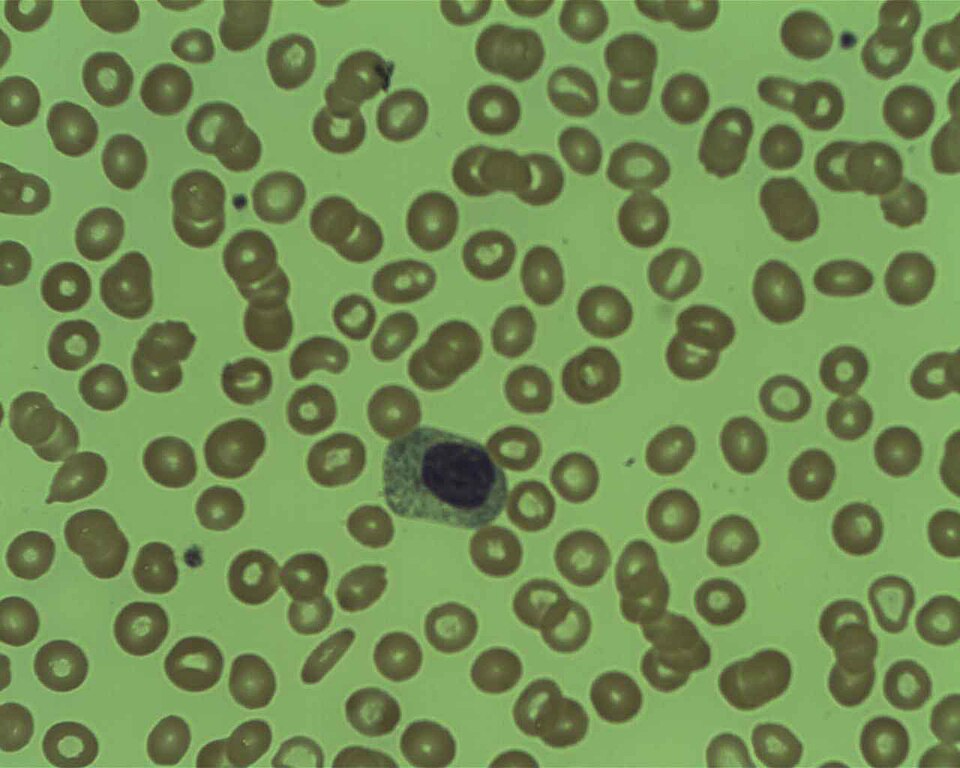
Myelocyte on bone marrow preparation, Giemsa stain. Normal morphology: round nucleus, pale cytoplasm.
Prof. Osaro Erhabor · CC BY-SA 4.0 · source
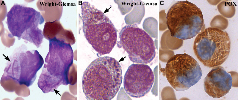
Acute promyelocytic leukemia (APL): Wright-Giemsa and MPO staining of promyeloblasts with Auer rods (arrows). The myelocyte's antecedent stage, arrested in APL.
Ru YX et al. Blood Sci 2023 (PMID 37228776) · CC BY 4.0 · source
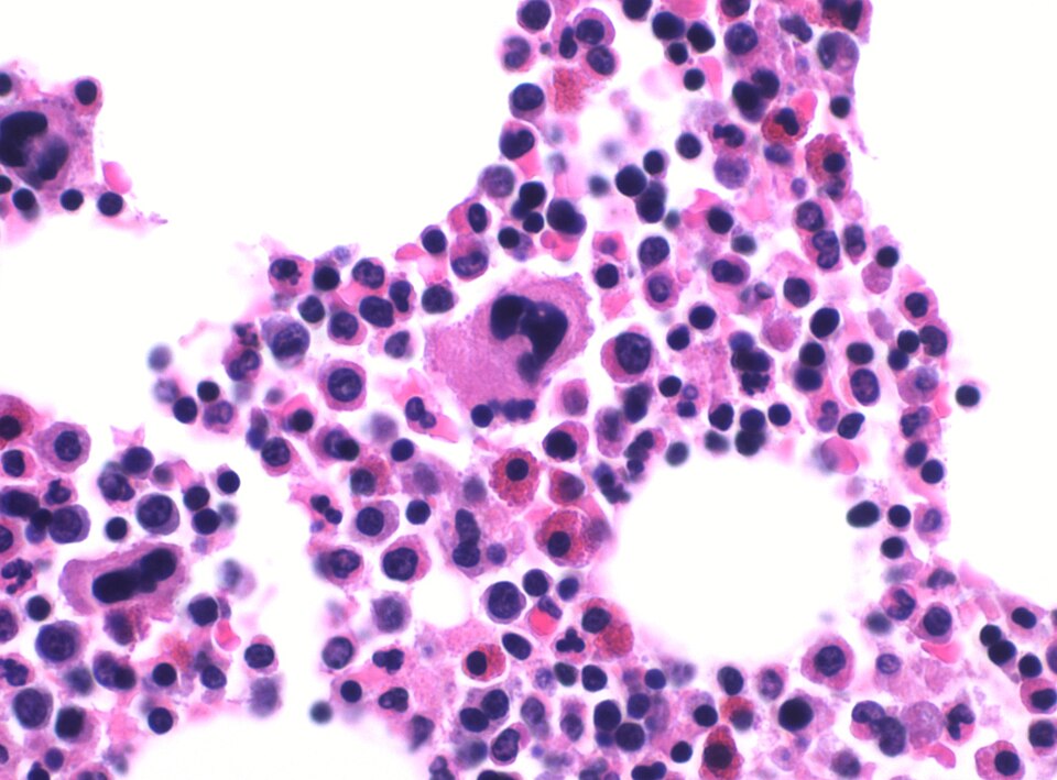
Mikael Haggstrom M.D. · CC0 · source
Illustrations
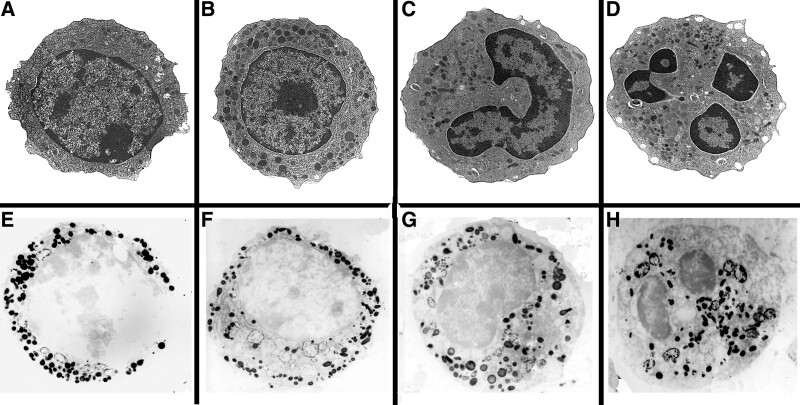
Normal neutrophils in (A)-(D), showing developmental stages of myeloblasts: myeloblasts (A), promyelocytes (B), myelocytes (C) to segmented granulocytes (D), ie, the mature neutrophils, x5k. (E)-(H) Positive myeloperoxidase reactivity of primary granules of the neutrophil stages from myeloblasts, promyelocytes. myelocytes to segmented granulocytes respectively, x5k.
Ru YX et al. Blood Sci 2023 (PMID 37228776) · CC BY 4.0 · source
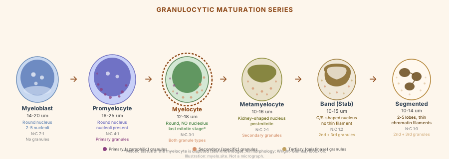
Granulocytic maturation series: myeloblast through segmented neutrophil, with nucleus shape, size range, granule types, and N:C ratio. The myelocyte stage (center, outlined).
myelo.site, after Bainton et al. 1971 · CC BY-SA 4.0 · source